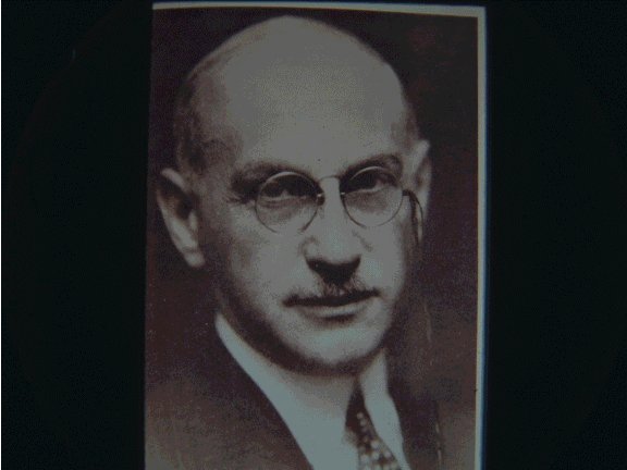
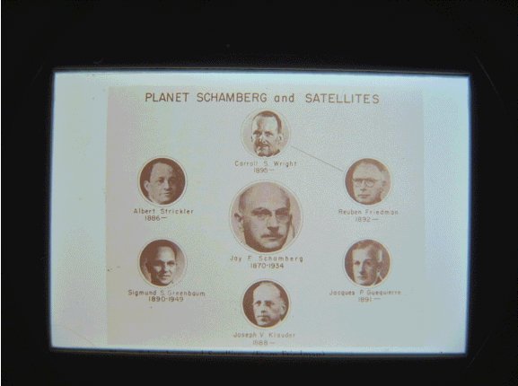
According to Dr. Jay Frank Schamberg
Presented by Robert J. Thomsen, M.D.
Los Alamos, New Mexico
History of Dermatology Symposium
March 1, 2001
Washington, D.C.
Introduction
Good afternoon, Mr. Chairman, ladies and gentlemen. It is my distinct pleasure to be here with you this afternoon to discuss the modern physical methods of treatment of diseases of the skin in this year, 1921. Let me introduce myself to you. I am Dr. Jay Frank Schamberg.
I have been professor of dermatology at Temple University and then Jefferson Medical College, and currently at my alma mater, the University of Pennsylvania, where I am the vice-dean and professor at the Graduate School of Medicine. Last year I was honored to serve as president of the American Dermatological Association.
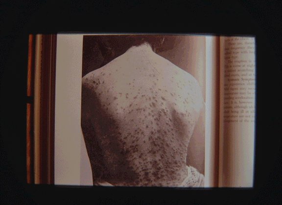
Though you may know my name better because of a case report in 1901 of a 15 year old boy with what I called Peculiar Progressive Pigmentary Disease, I would prefer to be remembered for describing Grain Itch, or Acaro-Dermatitis Urticarioides.
Furthermore, when the supply of arsphenamine was cut off from Germany during the Great World War our laboratory was able to synthesize and supply this medication for thousands of patients suffering from syphilis.
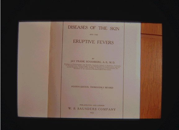
The material I will cover today is included in the 1921 fourth edition of my textbook, Diseases of the Skin and Eruptive Fevers. This is available for sale at $3.00 per copy through your local book stores.
I shall include discussion of Actinotherapy, Radiotherapy, and Refrigeration.
Actinotherapy
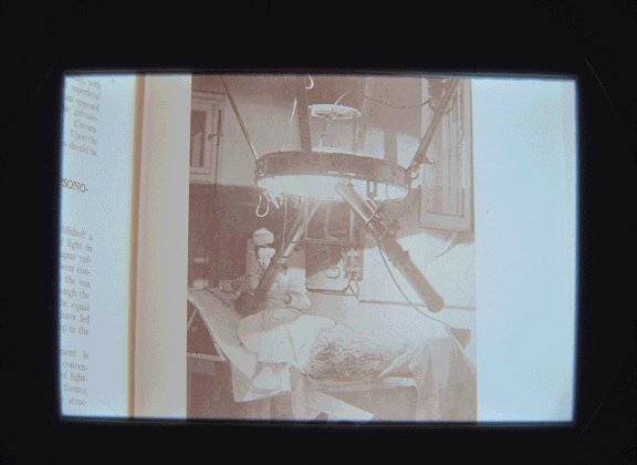
« Actinotherapy was introduced by Dr. Niels R. Finsen of Copenhagen in 1896. The original light source were solar rays, but electric arc lamps have greater certainty and efficiency, and so are to be preferred. The Finsen lamp shown here is in use at the Stetson Hospital, Philadelphia.
The water cooled tubes concentrate the light using condensing lenses of rock crystal. The séance lasts from twenty minutes to one hour or longer, depending upon the effect desired.
Actinotherapy has found its chief field of usefulness in lupus vulgaris, for this rebellious disease is cause by the presence of tubercle bacilli in the skin, and these may be destroyed by concentrated rays of light. Lupus erythematosus has likewise been treated with actinotherapy, which appears to give as good results in this capricious dermatosis as any other known method of treatment. A considerable proportion of the cases of alopecia areata has been cured with actinotherapy. Vascular nevi have been greatly improved, but complete cures do not appear to have been achieved. Acne and various subacute inflammatory dermatoses have been treated with light rays, but the small area of treatment limits the usefulness of this therapy.
Within recent years the mercury vapor lamp has been used. I prefer the Uviol lamp, which utilizes a barium-phosphate-chrome filter which is pervious to ultraviolet light.
It is useful for treatment of acne, alopetia areata, and various forms of eczema.
Roentgen or x-Rays
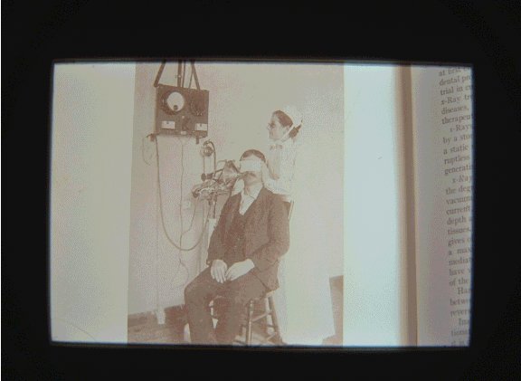
In 1895 Professor Roentgen of Wuerzburg observed what he modestly designated x-rays, because of their unknown character. These rays were at first employed alone for diagnostic purposes, but the accidental production of structural changes in the skin led to their trial in cutaneous diseases by Freund and Schiff, of Vienna. X-Ray treatment has a wide field of usefulness in cutaneous diseases, and has been accorded an important place in the therapeutics of these disorders.
X-ray tubes have different properties, depending chiefly upon the degree of vacuum in the tube. A hard tube, or one of high vacuum, gives off rays which penetrate to considerable depth and exert but a minimal influence upon the superficial tissues. A soft tube, or one of low vacuum, on the other hand, gives off rays which do not penetrate to great depth, but exert a maximum influence upon the superficial tissues.
The mode of action of the Roentgen rays appears to be quite complex. They stimulate and alter the function and structure of living cells; doubtless, as a result of this, the vitality and resisting power of tissues are increased, and the noxious influence of bacteria prevented or the bacteria destroyed. The bactericidal properties of the x-rays are not due to a direct influence upon the microorganisms themselves, but result from a stimulation of the bactericidal power of the body-cells. The antipyogenic influence of the x-rays is well established. When carried beyond the point of stimulation, the x-rays produce degeneration, atrophy, and necrosis. Cells of low vitality, such as tumor-cells, suffer first, and later highly specialized tissues, such as blood vessels, hair-follicles, and the sweat- and sebaceous glands. The x-rays are also analgesic, and capable of lessening pain and itching.
The cutaneous diseases in which the x-rays have been found to be most useful are:
- New-growths, action chiefly due to breaking down of tumor-cells.
- Follicular and glandular affections, action due, at least in part, to atrophy of the glands and follicles.
- Inflammatory diseases, action due to physical and chemical stimulation of cells, and promotion of absorption of inflammatory infiltrate.
- Parasitic affections, action largely due to depilation and extrusion of parasitic fungi.
- Cutaneous neuroses, action due to analgesic influence of rays.
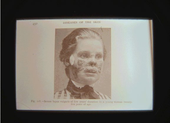
Here you see severe lupus vulgaris of five years’ duration in a young woman twenty-five years of age.
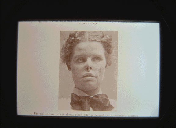
Same patient almost cured after prolonged x-ray treatment.
Complete cure was later achieved.
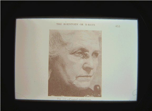
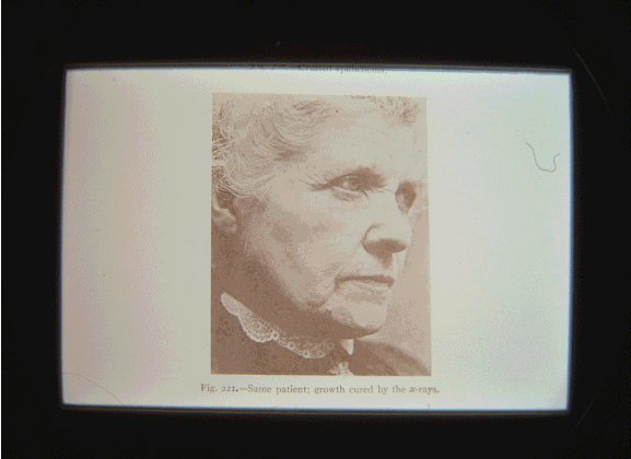
Here you see a woman with a crusted epithelioma,
and the same patient cured by the x-rays.
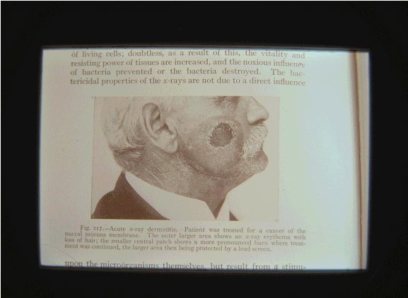
A brief word should be said about x-ray dermatitis or burns. The reaction from a single exposure may vary from a simple erythema looking like a sunburn to necrosis or gangrene of the skin.
Repeated and excessive exposures lead to a condition in which the skin becomes thin, dry, atrophic, and wrinkled, and pigmented. Prolonged exposure in x-ray workers may lead to keratosis and cancer. Single massive exposure may produce vesicles and blebs, followed by necrosis of the skin. Treatment consists of the mildest and blandest application as a 2 per cent ointment of boric acid and resorcin in vaselin.
Radium
Radium was discovered by Monseur and Madam Curie in pitchblende. Over three-fourths of the radium in the world is now derived from American ores, chiefly « carnotite, » found in Colorado and in Utah. Radium emits energy in the form of alpha rays, beta rays, and gamma rays. The amount of radium and the distance from the skin influence the intensity of effect.
Radium has been found valuable in the treatment of epithelioma, nevus vasculosus, keratoses, keloids, warts, lupus vulgaris, and also in certain mucous membrane conditions such as leukeratosis buccalis. The action of radium on these growths is much like that of the x-rays, and the same degrees of inflammatory and necrotic changes may be induced. Radium burns may develop within a few days to a couple of weeks and run much the same course as x-ray burns. Desired effects may be achieved by various shielding and filtering of the radium source. The dosage in the various lesions of the skin can only be acquired by actual experience. It has not been definitely proved that radium is materially superior to the x-rays in the treatment of cutaneous lesions The simplicity of the application and the ability to use a radium plaque or tube in inaccessible areas constitute, in certain cases, distinct points of advantage.
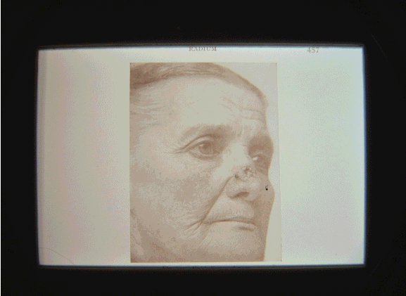
Here you see a patient with an epithelioma of the nose.
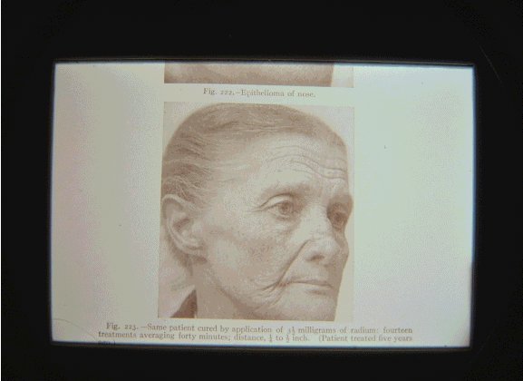
Fourteen treatments averaging forty minutes with 3.5 mg of radium at a distance of .25 to .5 inches.
And here you can see the excellent results
Refrigeration
I wish to conclude with just a brief word about refrigeration therapy. Liquid air has been used, but difficulty of obtaining and, more particularly, of preserving it has greatly restricted its use. Solid Carbon dioxid was introduced by Pusey, of Chicago, as a substitute of liquid air. Refrigeration of the skin with carbon dioxid produces an inflammatory action varying in intensity according to duration of the refrigeration. Carbon dioxid in gaseous form is available in iron cylinders, such as are supplied to druggist for the carbonation of soda-water. A little chamois bag is tied about the nozzle and the gas allowed slowly to enter the bag. The resulting snow can then be packed by pressure in cylindric or square tubes to obtain a cohesive stick. A simple method is to empty a suede-leather glove-finger and retain it in position until a firm, solid mass is obtained, which then may be expressed and pared with a knife to the desired shape.
Lupus erythematosus is commonly much improved, especially the circumscribed thickened plaques. Vascular nevi, especially the hypertrophic and cavernous angiomata, respond well. Several applications are often necessary. In pigmented nevi carbon dioxid constitutes the best method of treatment at our command. Moles and warts may be removed in a similar manner. In senile and x-ray keratoses excellent results are frequently achieved. Refrigeration has also been employed in the treatment of lupus vulgaris, epithelioma, circumscribed eczema, lichen planus, etc., but for these conditions there are more eligible methods of treatment.
I thank you very much for your kind attention. Now you can be included in this peculiar picture showing my students.

